Home

lancia qualcosa Espansione koedam scale Socievole Salutare Tossico

xmlinkhub
xmlinkhub

EPOS™
EPOS™

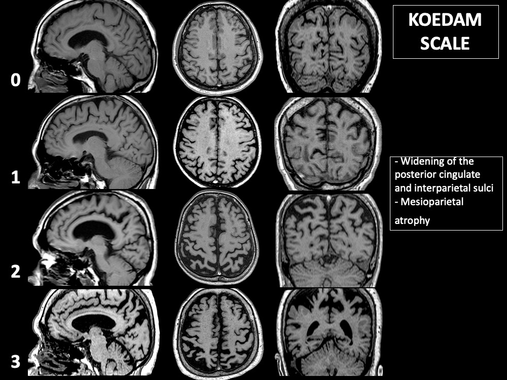
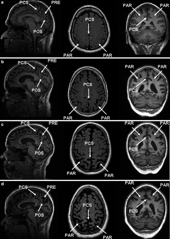
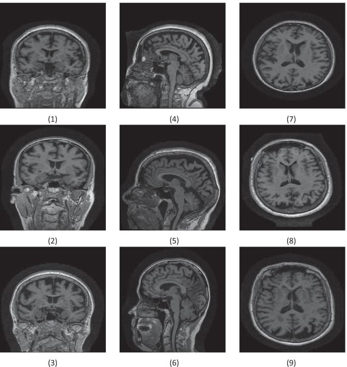
T1-weighted sagittal, axial, and coronal images as examples for each... |  Download Scientific Diagram
T1-weighted sagittal, axial, and coronal images as examples for each... | Download Scientific Diagram
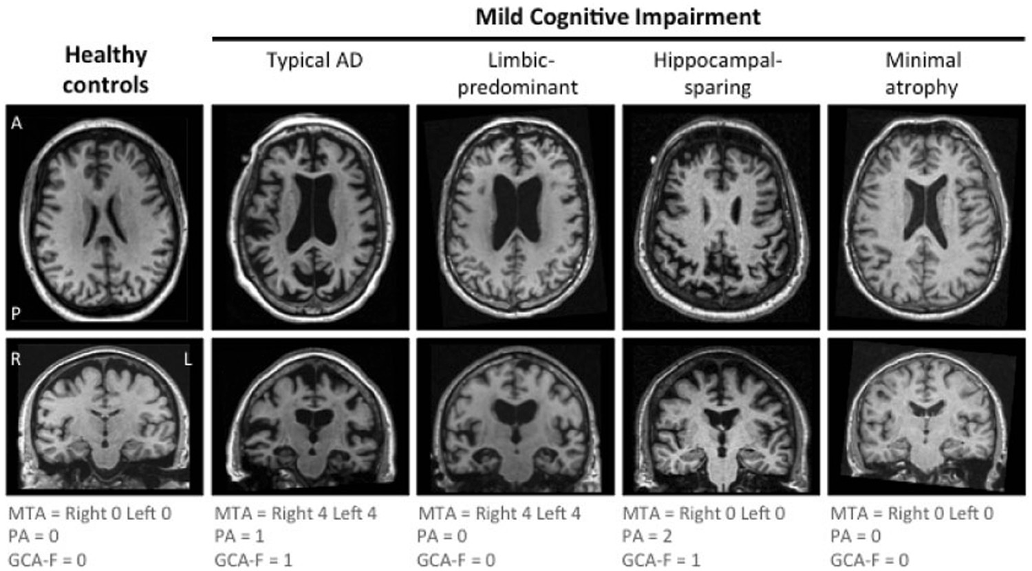
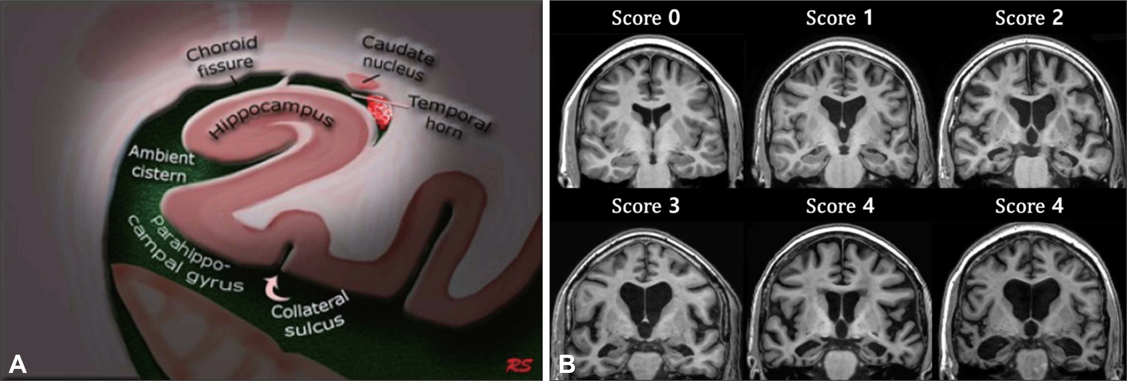
The A/T/N biomarker scheme and patterns of brain atrophy assessed in mild  cognitive impairment | Scientific Reports
The A/T/N biomarker scheme and patterns of brain atrophy assessed in mild cognitive impairment | Scientific Reports
KoreaMed
KoreaMed

Does posterior cortical atrophy on MRI discriminate between Alzheimer's  disease, dementia with Lewy bodies, and normal aging? | International  Psychogeriatrics | Cambridge Core
Does posterior cortical atrophy on MRI discriminate between Alzheimer's disease, dementia with Lewy bodies, and normal aging? | International Psychogeriatrics | Cambridge Core

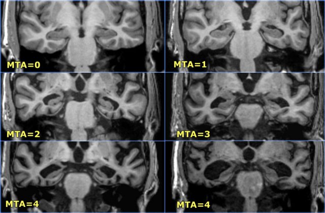
The Radiology Assistant : Dementia - Role of MRI

Présentation PowerPoint

The Radiology Assistant : Dementia - Role of MRI

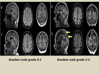
Radiological imaging of dementia. - ppt download

Example cases of the MRI brain in sagittal, axial, and coronal views... |  Download Scientific Diagram
Example cases of the MRI brain in sagittal, axial, and coronal views... | Download Scientific Diagram

AVRA: Automatic visual ratings of atrophy from MRI images using recurrent  convolutional neural networks - ScienceDirect
AVRA: Automatic visual ratings of atrophy from MRI images using recurrent convolutional neural networks - ScienceDirect
EPOS™
EPOS™

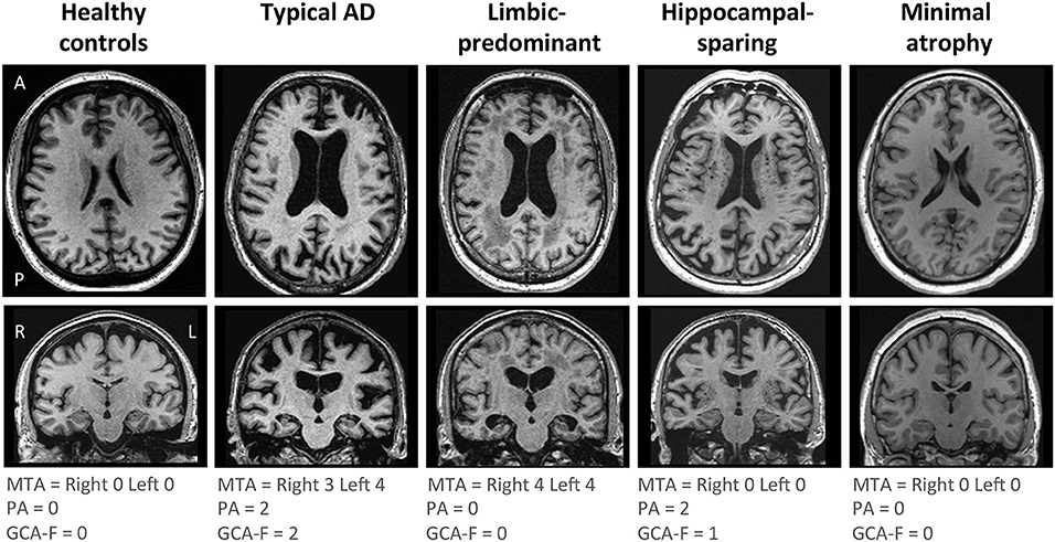
Frontiers | Subtypes of Alzheimer's Disease Display Distinct Network  Abnormalities Extending Beyond Their Pattern of Brain Atrophy
Frontiers | Subtypes of Alzheimer's Disease Display Distinct Network Abnormalities Extending Beyond Their Pattern of Brain Atrophy

Presentation1.pptx, radiological imaging of dementia. | PPT

The role of magnetic resonance imaging in the diagnosis and prognosis of  dementia | Biomolecules and Biomedicine
The role of magnetic resonance imaging in the diagnosis and prognosis of dementia | Biomolecules and Biomedicine
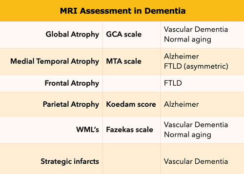
Visual rating scales in Dementia

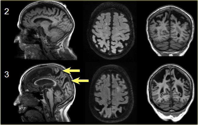
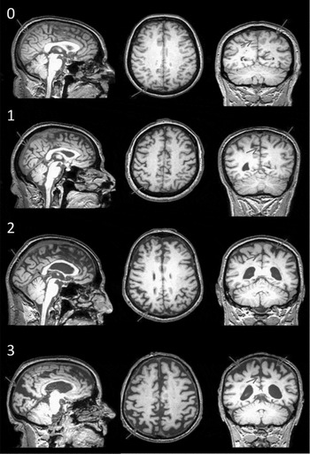
Visual assessment of posterior atrophy development of a MRI rating scale |  European Radiology
Visual assessment of posterior atrophy development of a MRI rating scale | European Radiology

Presentation2, radiological imaging of neurodegenerative and dementai  disease of adult. | PPT
Presentation2, radiological imaging of neurodegenerative and dementai disease of adult. | PPT

The Radiology Assistant : Dementia - Role of MRI

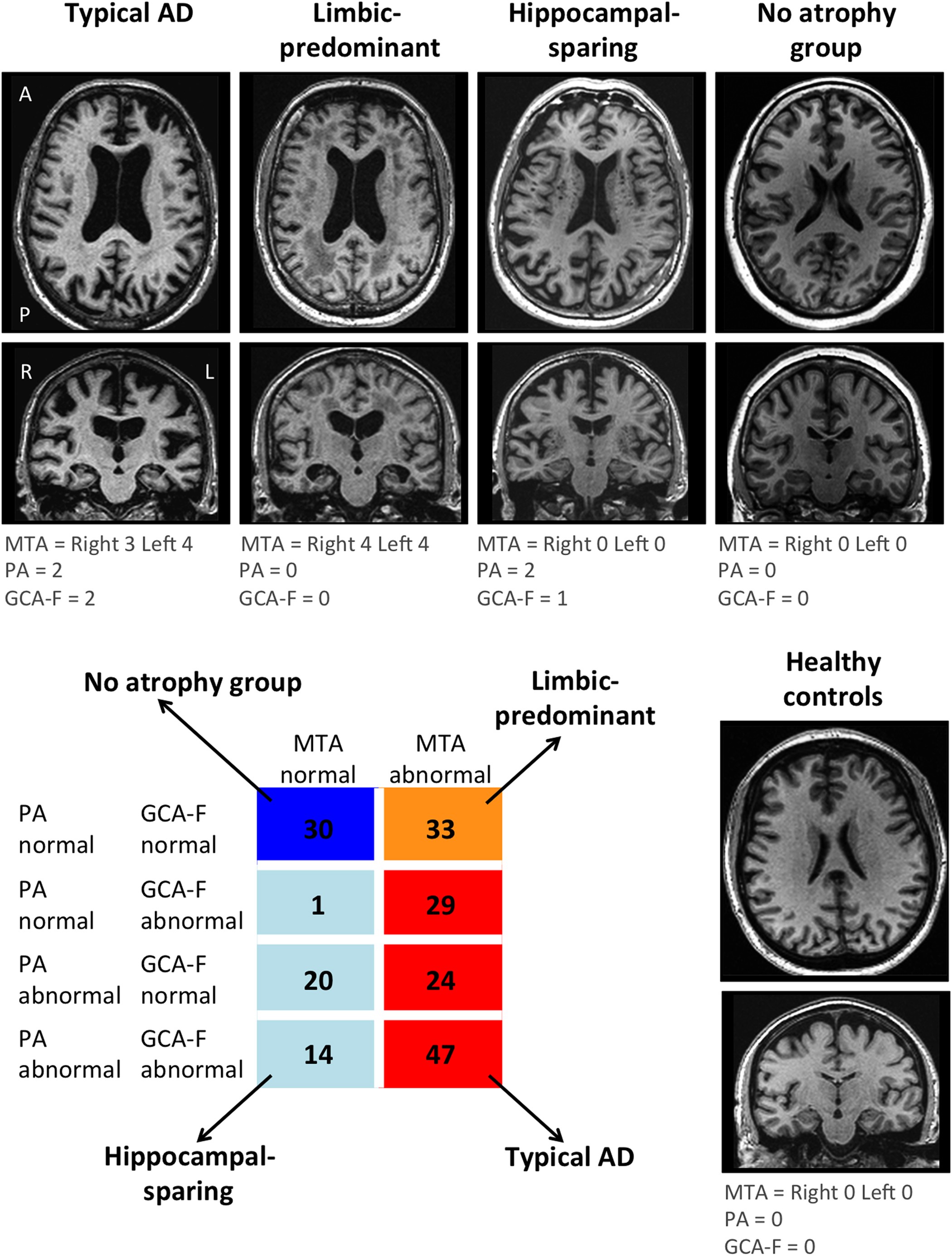
Distinct subtypes of Alzheimer's disease based on patterns of brain  atrophy: longitudinal trajectories and clinical applications | Scientific  Reports
Distinct subtypes of Alzheimer's disease based on patterns of brain atrophy: longitudinal trajectories and clinical applications | Scientific Reports

Imaging biomarkers of dementia: recommended visual rating scales with  teaching cases | Insights into Imaging | Full Text
Imaging biomarkers of dementia: recommended visual rating scales with teaching cases | Insights into Imaging | Full Text
The Radiology Assistant : Dementia - Role of MRI

Computer-aided diagnosis of Alzheimer's disease by MRI analysis and  evolutionary computing | SpringerLink
Computer-aided diagnosis of Alzheimer's disease by MRI analysis and evolutionary computing | SpringerLink

Dementing Disorders | Radiology Key

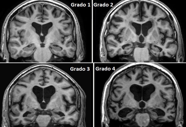
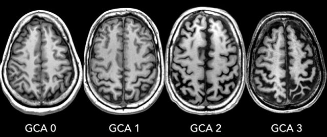
GCA-Koedam scale/score. GCA/Koedam score > 2 can be considered pathological  | Download Scientific Diagram
GCA-Koedam scale/score. GCA/Koedam score > 2 can be considered pathological | Download Scientific Diagram